Published in PLOS One on 2026-09-29, this study is presented as an attributed evidence briefing. The excerpts below are the original researchers’ words, reproduced under their Creative Commons Attribution licence. HealthSignal has selected the abstract and, where needed, opening background paragraphs; the complete methods, results, limitations and references remain in the linked paper.
Original authors: Michelle Gellhorn Serra, Cornelius Rohde, Lucie Sauerhering, Lars Meier, Lennart Kämper, Pauline Neubecker, Markus Eickmann, Alexandra Kupke, Stephan Becker, Anke-Dorothee Werner
Source: PLOS One • DOI 10.1371/journal.pone.0347080
Read the complete primary study ↗ · Creative Commons Attribution licence
From the researchers
Abstract
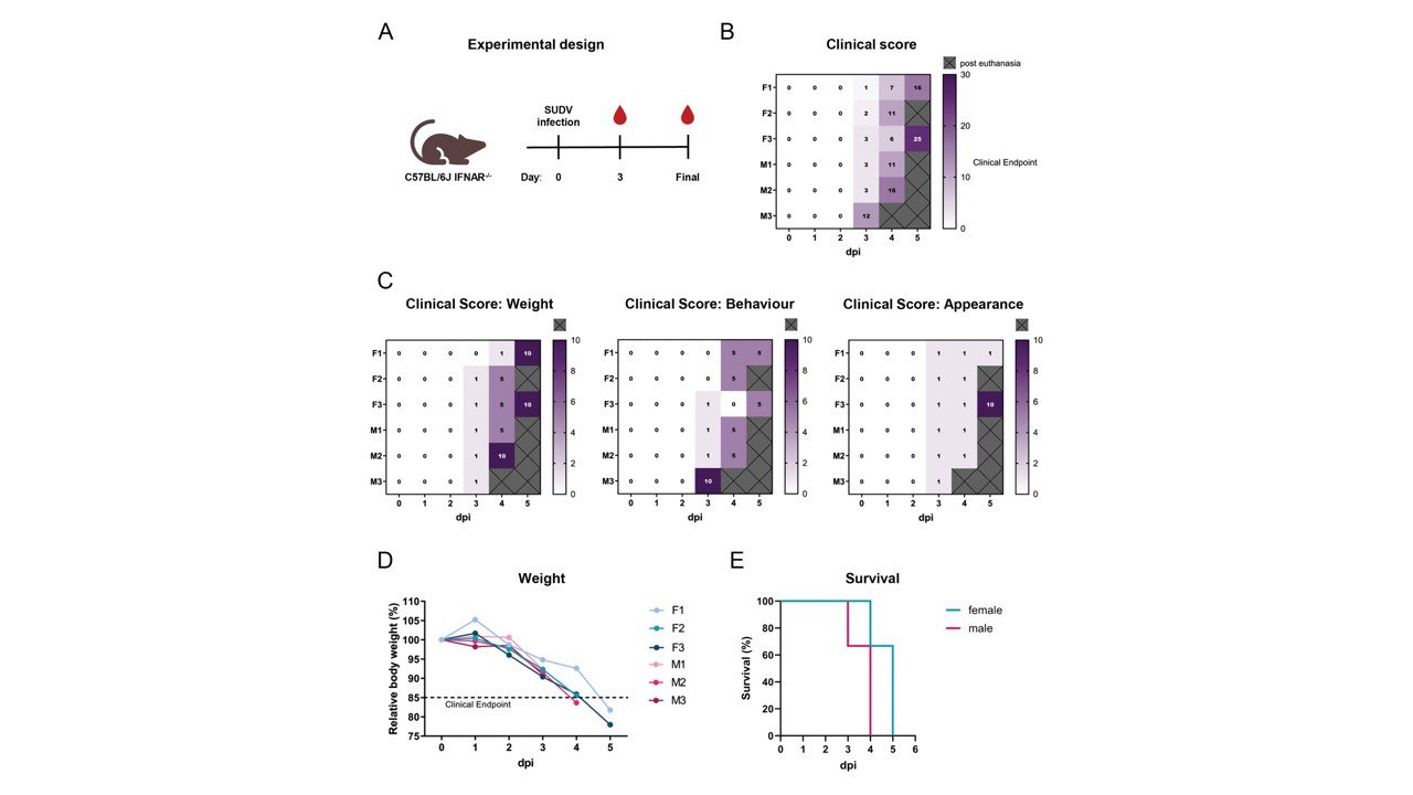
Sudan virus (SUDV) is a member of the family Filoviridae, which comprises highly pathogenic viruses associated with unusually high case fatality rates. The development of medical countermeasures against filoviruses, including antivirals, vaccines, and therapeutic antibodies, requires preclinical evaluation in suitable animal models. C57BL/6J IFNAR-/- mice, which lack the type I interferon (IFN-α/β) receptor, have been reported to be susceptible to filovirus infections, although their impaired innate immune response may represent a potential limitation of the model. Here, we characterized the clinical, virological, pathological, and inflammatory responses following SUDV Boniface infection in IFNAR-/- mice. Following infection, animals developed progressive clinical disease characterized by substantial weight loss and pronounced changes in behavior and appearance. Mice reached the predefined clinical endpoint 3–5 days post infection. Post mortem analysis of terminal samples revealed high viral loads and viral genome copies in all tested organs as well as in serum, consistent with widespread systemic dissemination. Histopathological examination revealed prominent lesions, particularly in the liver and spleen. In addition, terminal serum analysis revealed a broad and pronounced inflammatory response, with significantly elevated concentrations of multiple cytokines and chemokines compared with non-infected controls, encompassing pro-inflammatory, regulatory, and hematopoietic mediators. Together, these findings characterize the clinical course, systemic viral dissemination, tissue pathology, and terminal inflammatory response associated with lethal SUDV infection in IFNAR-/- mice. The observed cytokine and chemokine elevations are consistent with inflammatory responses described in filovirus infections, although the single terminal sampling time point and limited cohort size preclude conclusions regarding the temporal development or mechanistic basis of this response. These data support the utility of this model for investigating filovirus pathogenesis and infection-associated immune responses and warrant further evaluation of its suitability for SUDV infection and for preclinical studies of filovirus countermeasures.
Introduction
The Sudan virus (SUDV) belongs to the order Mononegavirales and is a member of the genus Orthoebolavirus within the Filoviridae family. Since SUDV was first reported in 1976, the virus has periodically emerged, causing nine additional outbreaks with case fatality rates ranging between 41 and 70% [1,2]. SUDV and other filoviruses are listed as priority pathogens by the WHO R&D Blue-print initiative as they pose a great threat to public health care systems because of their epidemic potential and insufficient medical countermeasures [3]. Most filoviruses are therefore classified as biosafety level-4 (BSL-4) pathogens.
Wild-type (WT) rodents are not susceptible to filoviruses and show minimal or no symptoms. After adaptation through serial passaging in rodents, filoviruses adopt genomic mutations which are accompanied by the development of severe disease [4–6]. As a result, these adapted viruses may not accurately reflect WT pathogenesis and vaccines, antibodies or small molecule antivirals developed against adapted viruses might not be optimal to counteract WT virus infection. Immunocompromised rodents therefore represent a useful tool that supports WT filovirus infection efficiently [4,7,8]. Common rodent models include the interferon-α/β receptor knockout mice (IFNAR-/-), the double interferon-α/β and γ receptor knockout (IFNAGR-/-), interferon-γ receptor knockout (IFNGR-/-), the cytoplasmic signal transducer and activator of transcription-1 protein knockout (STAT-1-/-) or the severe combined immunodeficiency (SCID) mice. Their inherently compromised immune responses represent limitations regarding infection and disease progression, as detailed information on the mounted immune response in successfully infected animals is missing for a number of mouse and virus strain combinations. Nevertheless, IFNAR-/- mice, for instance, can efficiently mount humoral and cellular immune responses, and are therefore commonly used to evaluate different vaccine strategies, including those for Zika virus [9], Ebola virus [8,10,11] and Crimean-Congo haemorrhagic fever virus [12]. For SUDV, several of such immunocompromised mouse models exist with different combinations of mice and virus strains [4,7,8,13–17]. The lethality of SUDV infection in these models varies and the majority of these studies focused on haematology and serum chemistry. This variability is likely driven by differences in viral isolates, experimental conditions, and humane endpoint criteria, which together influence the observed disease severity and lethality.
Reading this evidence
Use the full paper to check the study design, participant selection, comparator and outcome definitions before drawing a practical conclusion. An association does not by itself show cause and effect. A reported performance measure needs its denominator and uncertainty. A finding from a particular facility, sample or time period should retain that scope when it is discussed elsewhere.
This briefing makes the source accessible within the wider public health desk. It does not assess an individual patient or certify a product. When considering relevance to another health system, examine the original authors’ account of context and limitations, then seek local implementation evidence. The related reporting below provides other studies to read alongside this one; it is a topic connection, rather than a claim that their results are directly comparable.
© 2026 Michelle Gellhorn Serra, Cornelius Rohde, Lucie Sauerhering, Lars Meier, Lennart Kämper, Pauline Neubecker, Markus Eickmann, Alexandra Kupke, Stephan Becker, Anke-Dorothee Werner. Excerpts reproduced under CC BY. Selection, formatting and contextual notes by HealthSignal. The researchers are the authors of the source study, not members of HealthSignal’s staff.
Explore connected evidence
- Evidence brief: Serological responses to SARS-CoV-2 in urban and rural Ghana: Antibody waning and implications for long-term population immunity
- Evidence brief: Development and qualification of an enzyme-linked immunosorbent assay to detect human serum immunoglobulin G reactive to multiple lineages of Lassa virus nucleoprotein
- Evidence brief: Tuberculosis-related knowledge, attitudes, and self-reported preventive practices among medical students at the University of Khartoum: A cross-sectional educational survey