Published in PLOS One on 2026-02-06, this study is presented as an attributed evidence briefing. The excerpts below are the original researchers’ words, reproduced under their Creative Commons Attribution licence. Health Signal has selected the abstract and, where needed, opening background paragraphs; the complete methods, results, limitations and references remain in the linked paper.
Original study title: Sex hormone profiles and associated factors among adult tuberculosis patients at Gondar town, northwest Ethiopia: A comparative cross-sectional study
Original authors: Eshet Gebrie, Habtamu Wondifraw Baynes, Berihun Agegn Mengistie, Temesgen Kassie, Zeleke Kassahun, Abebe Birhanu, Amanuale Zayede, Elias Chane
Source: PLOS One • DOI 10.1371/journal.pone.0340631
Read the complete primary study ↗ · Creative Commons Attribution licence
From the researchers
Abstract
Tuberculosis, caused by Mycobacterium tuberculosis, is the second leading cause of death from infectious diseases worldwide. Tuberculosis is associated with alterations in sex hormone levels, particularly testosterone, estradiol, and progesterone. However, previous studies have reported conflicting results, with some showing increased or decreased levels in tuberculosis positive patients, while others found no significant differences. This study aims to assess and compare sex hormone profiles among adult tuberculosis-positive patients and tuberculosis-negative individuals and to identify associated factors.
A comparative cross-sectional study was conducted from June 15 to August 20, 2024, among 300 eligible adult tuberculosis-positive patients and age-matched tuberculosis-negative individuals in five selected health institutions in Gondar town. Participants were recruited using a simple random sampling technique, and sociodemographic, clinical, anthropometric, and behavioral data were collected through a structured questionnaire. Five milliliters of venous blood were used to determine hormone levels using the Beckman Coulter DXI 800 chemistry hormonal analyzer. Hypogonadism was defined by sex-specific hormones and categorized as primary, secondary, and subclinical. The data were analyzed using SPSS version 25.0. Descriptive statistics, independent t-tests, one-way ANOVA, Mann-Whitney U, Kruskal-Wallis H test, and bivariable and multivariable statistical models were used. A p-value < 0.05 with a 95% CI was considered statistically significant.
Male tuberculosis-positive patients showed significantly higher estradiol, luteinizing hormone, and FSH (p < 0.001), but lower testosterone (p < 0.001). Newly diagnosed tuberculosis-positive patients had significantly lower progesterone levels (p < 0.005). Female tuberculosis-positive patients showed significantly lower testosterone and progesterone but higher follicle-stimulating hormone levels compared to tuberculosis-negative individuals (P < 0.001). Estradiol and luteinizing hormone levels did not differ significantly in female tuberculosis-positive patients. However, newly diagnosed tuberculosis-positive patients had significantly higher median estradiol levels (p < 0.001). The overall prevalence of hypogonadism was 30.3% (95% CI (25.2–35.9%)), sex [AOR = 11.36, 95% CI (3.6, 36.17)] (p < 0.001), dietary diversity (participants with lower diversity, including those with no dietary diversity [AOR = 8.98, 95% CI (2.37, 33.99)] (p = 0.001), those with sometimes [AOR = 9.2, 95% CI (2.77, 30.62)] (p < 0.001) and a usual dietary diversity [AOR = 3.24, (1.04, 10.06) (p = 0.042), and cortisol [AOR = 4.01, 95% CI (1.7, 9.5)] (p = 0.002) levels were significant determinants of hypogonadism in tuberculosis patients.
Male tuberculosis-positive patients showed significantly higher mean estradiol, luteinizing hormone, and follicle-stimulating hormone but lower testosterone levels, while female tuberculosis-positive patients had significantly lower testosterone and progesterone and higher follicle-stimulating hormone levels. There was a higher prevalence of hypogonadism, particularly in males. Early diagnosis of these hormonal changes is crucial in tuberculosis-positive patients to prevent complications such as impaired gonadal function related to hormonal disturbances.
Reading this evidence
Use the full paper to check the study design, participant selection, comparator and outcome definitions before drawing a practical conclusion. An association does not by itself show cause and effect. A reported performance measure needs its denominator and uncertainty. A finding from a particular facility, sample or time period should retain that scope when it is discussed elsewhere.
This briefing makes the source accessible within the wider research & evidence desk. It does not assess an individual patient or certify a product. When considering relevance to another health system, examine the original authors’ account of context and limitations, then seek local implementation evidence. The related reporting below provides other studies to read alongside this one; it is a topic connection, rather than a claim that their results are directly comparable.
© 2026 Eshet Gebrie, Habtamu Wondifraw Baynes, Berihun Agegn Mengistie, Temesgen Kassie, Zeleke Kassahun, Abebe Birhanu, Amanuale Zayede, Elias Chane. Excerpts reproduced under CC BY. Selection, formatting and contextual notes by Health Signal. The researchers are the authors of the source study, not members of Health Signal’s staff.
Explore connected evidence
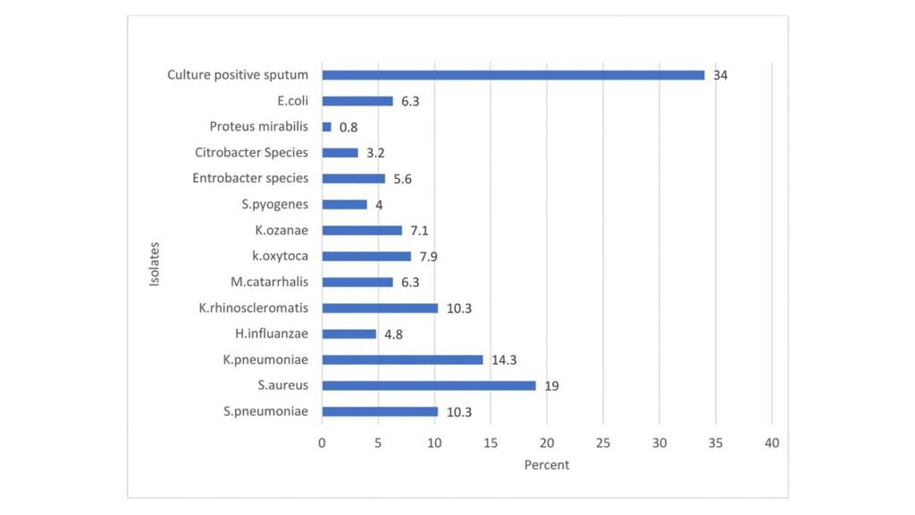
- Evidence brief: Uncovering overlooked bacterial infections in gene X-pert MTB/RIF negative sputum samples of adult patients with presumptive tuberculosis in Harar, Eastern Ethiopia
- Evidence brief: Time to sputum culture conversion, treatment outcomes, and associated factors among rifampicin-resistant or multidrug-resistant tuberculosis patients in the Sidama region, Ethiopia: A retrospective follow-up study
- Evidence brief: Magnitude and predictors of anemia among adult solid cancer patients in Hawassa, Ethiopia: A cross-sectional study